Yolk Sac Herniation in Reptiles
5748 Views |

Yolk Sac Herniation in Reptiles
By Asst. Prof. Somphoth Weerakul, DVM, PhD (Dr. Kaew)
In many clinical case studies, we often observe internal organ-like tissues protruding from the abdominal wall at the site of the umbilicus (navel). Naturally, no internal organs should remain outside the body except for a small portion of the yolk sac. In a healthy animal, these organs are contained within the celomic cavity, and the inner abdominal wall should be sealed. While the skin layer may remain open initially, it gradually closes over time—ranging from a few weeks to two months—depending on the species, breed, incubation temperature, nutrition, growth rate, and overall body condition.
Yolk Sac Herniation is a common issue encountered in reptiles, including snakes, lizards, turtles, and crocodiles. It is frequently seen in Corn Snakes, Ball Pythons, Crocodilians, and Iguanas, and is also a regular occurrence in birds.
Risk Factors and Physiology
It is widely understood that high humidity and prolonged incubation temperatures are significant risk factors. When temperatures are lower than the optimal range for embryonic development, the incubation period is extended. Adjusting the temperature to the appropriate higher level can help reduce these occurrences. However, other primary risk factors exist.
The yolk sac serves as the vital nutrient source for the embryo. It is located between the jejunum and ileum of the small intestine, connected by the vitelline duct. This duct transports the non-soluble fraction of the yolk into the small intestine for absorption, while the soluble fraction is absorbed directly into the bloodstream. After hatching, the remaining yolk continues to provide nourishment for 1–2 weeks, allowing neonates time to develop hunting instincts before they begin feeding independently.
Causes of the Problem
Beyond low incubation temperatures, studies show two other major causes:
Eggshell Contamination: Infection originating from the shell before hatching.
Maternal Transmission: Direct transmission from the mother to the embryonic membranes.
These infections lead to omphalitis (inflammation of the navel), which spreads to the yolk sac and eventually into the celomic cavity. This can result in enteritis (inflammation of the intestines), peritonitis, and internal organ failure, leading to septicemia (blood poisoning).
Common pathogens found in reptiles include Pseudomonas aeruginosa, Aeromonas hydrophila, Edwardsiella spp., and Klebsiella spp. A particularly critical pathogen is Salmonella spp., specifically the serotype S. arizonae. My colleagues (Dr. Wichit and Ms. Paweena, a medical technologist) and I conducted a random survey across four regions of Thailand and found that exotic snakes kept in Thailand have a higher prevalence of this bacteria compared to turtles and lizards.
Once the yolk sac becomes infected, fibrin or connective tissue forms around it, hindering or completely failing the absorption of yolk into the bloodstream. This often causes the vitelline duct to constrict, trapping the yolk inside the sac. Consequently, the yolk sac remains enlarged both internally and externally. Because the yolk is an excellent nutrient source for bacteria, it turns into a cream-like consistency or thick pus. The toxins produced by these bacteria are a primary cause of neonatal mortality.
In mild cases of yolk retention or herniation, the hatchling may exhibit anorexia (loss of appetite), weight loss, and stunted growth. Without prompt intervention, these animals often fail to thrive or die.
Preventive Measures
Optimal Incubation: Thoroughly study the specific temperature and humidity requirements for each species. In practice, adjustments based on the specific clutch or mother may be necessary.
Shell Hygiene: If the problem recurs in your collection, consider culturing bacteria from the eggshells or cleaning eggs before incubation to reduce contamination. Some use plain water, while others prefer mild disinfectants.
Incubator Sanitization: Clean and disinfect incubators before use. Options include:
Abreuval (Hydrogen peroxide + Biguanide hydrochloride): Often used in livestock; safe and can be evaporated within the incubator at normal temperatures ($32$°C).
F10 (Benzalkonium chloride + Polyhexamethylene biguanide): Popular in crocodile and bird farms.
Chlorhexidine: Also a viable option for surface cleaning.
Post-Hatch Care: Since the navel is not yet closed at birth, hatchlings can be dipped in or wiped with diluted disinfectants (e.g., Quaternary ammonium or F10). In some cases, the umbilical stalk may be ligated or sutured and treated with Povidone-iodine.
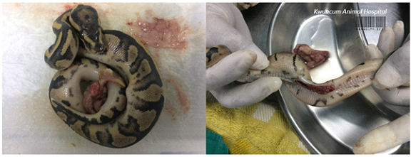
Treatment of Yolk Sac Herniation
When a herniated yolk sac is present, extreme caution regarding infection is required. Contamination can occur post-hatching from the environment because the navel is still open.
Surgical Intervention: Surgical removal of the yolk sac or suturing the navel is often necessary, especially if the opening is wide and communicates with the celomic cavity.
Examination: Even if the herniation appears minor, ensure that it is only the yolk sac. In many cases, a wide umbilical opening may pull the attached small intestine out as well.
Procedure: The yolk sac should be ligated at the vitelline duct, separating it from the small intestine before removal.
Debridement: Check for inflammation of surrounding organs. If necrotic (dead) tissue is found, it must be debrided and cleaned before returning the organs to the cavity and suturing the skin.
Follow-up: If infection is already present, the animal must receive ongoing medical treatment until fully recovered.